Normal View Dyslexic View
Personalised surgery for the colon cancer patients: new frontiers
6 July 2022
Guest Blog Lower GI
Related articles

Low presence of intraluminal cancer cells in rectal washout during transanal total mesorectal excision.
Sharaf Karim Perdawood¹, Rebecca Svensson Neufert², Jens Kroeigaard¹, Pierre Jean-Claude Maina¹, Susanne Eiholm³, Fredrik Jörgren², Pamela Buchwald4
¹Department of Gastrointestinal Surgery, Slagelse Hospital, Denmark
²Department of Surgery, Helsingborg Hospital, Helsingborg, Lund University, Lund, Sweden
³Department of Histopathology, Region Zealand, Denmark
4Department of Surgery, Skåne University Hospital, Malmö, Lund University, Lund, Sweden
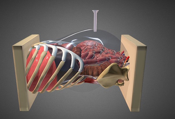
Local recurrence (LR) after rectal cancer surgery is a disastrous event occurring in up to 10% after transanal total mesorectal excision (TaTME) 1-3. LR usually arises within two years of primary surgery and is associated with significant morbidity 4. The adoption of total mesorectal excision (TME) has dramatically improved LR rates 5, although some studies have indicated early multifocal pelvic LR after TaTME 6, 7. Factors associated with LR are mainly related to non-radical surgery, intraoperative bowel perforation, and TNM stage 8-10. Another potential risk factor is the direct implantation of viable intraluminal cancer cells in the peri-anastomotic area 11-13. Different technical aspects, including rectal washout to eradicate free intraluminal cancer cells, may be important to achieve the best possible outcomes 14-18.
In recent years, TaTME was introduced as “a new solution to some old problems” 19. The procedure allows for precise visualisation of the operative field in the lowermost part of the pelvis 20. Rectal washout is an integrated part of TaTME. However, details about the procedure including the appropriate fluid type and volume, are not standardised 12.
Our recent pilot study examined fluid samples from rectal washout to determine the appropriate fluid volume needed to eliminate intraluminal cancer cells during TaTME. We hypothesised that since rectal washout during TaTME is performed after the placement of a snog purse-string suture without touching the tumour, it has a lower yield of cancer cells than rectal washout in normal TME dissection.
Author’s journey to extraperitoneal left colon surgery.
Tarek S Hany Department of Colorectal Surgery Lancashire Teaching NHS Foundation Trust Preston United Kingdom
From idea to practice.
The transperitoneal (TP) approach to colorectal surgery (both laparoscopic and robotic) has never made sense to me even after many years of performing both techniques. From a colorectal perspective, every important anatomical structure is a retroperitoneal structure. The peritoneum itself is an under-appreciated, under-utilised organ. When trespassing in the peritoneal cavity, the small bowel and omentum present significant unnecessary challenges especially in the obese and those with previous surgery. The whole experience could sometimes feel like trying a knife and a fork for the first time.
Multiport wristed robotic instruments have just made an awkward laparoscopic TP procedure less so. There is a design fault with TP access. There is no doubt, however, that the TP access works in many straightforward operations and this piece is not in any way a call to abandon TP access. However, some 10-25% of operations are responsible for 75-90% of a challenging, long, and painful operation (for patient, surgeon, assistant, and anaesthetist).
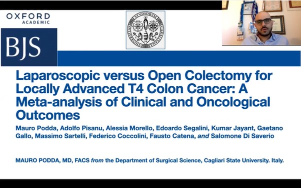
Laparoscopic versus open colectomy for locally advanced T4 colon cancer: a meta-analysis of clinical and oncological outcomes.
Mauro Podda MD FACS, Adolfo Pisanu MD PhD, Alessia Morello MD, Edoardo Segalini MD, Kumar Jayant MD PhD FACS, Gaetano Gallo MD, Massimo Sartelli MD, Federico Coccolini MD, Fausto Catena MD PhD FRCS, and Salomone Di Saverio MD FACS FRCS.
Colon cancer accounts for 9.4% and 10.6% of all tumours in the female and male populations, respectively [Sung H 2020]. Approximately 10-20% of patients diagnosed with colon cancer present with locally advanced tumours penetrating the surface of the peritoneum or directly invading other organs [Brenner H 2014].
This meta-analysis aimed to review the currently available evidence on laparoscopic colon cancer resection, evaluate short postoperative and long-term oncological outcomes after laparoscopic colectomy, and compare the above with conventional open surgery.
We searched medical databases for publications of comparative studies, including randomised controlled trials, prospective cohort studies, and retrospective cohort studies comparing laparoscopic and open surgery as a treatment for T4 locally advanced tumours colon cancers. The searches are up-to-date to April 2021.
Thank you to our strategic partners


.png)


Connect