BJS Academy>Cutting edge blog>Visual abstract: Mer...
Visual abstract: Merged SLEEVEPASS and SM-BOSS trials
1 February 2021
Guest Blog Upper GI
Related articles

Management of Crohn’s Disease. The shiny medical sports car or the worn out surgical banger?
Professor Steven R Brown, Sheffield Teaching Hospitals. Based on the BJS Lecture at ACPGBI 2020
Professor Steven R Brown, Sheffield Teaching Hospitals.
Based on the BJS Lecture at ACPGBI 2020
A success story
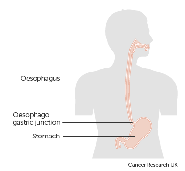
Guest blog: Keyhole versus open surgery for oesophageal cancer
B. P. Müller-Stich, P. Probst, H. Nienhüser, S. Fazeli, J. Senft, E. Kalkum, P. Heger, R. Warschkow, F. Nickel, A.T. Billeter, P. P. Grimminger, C. Gutschow, T. S. Dabakuyo-Yonli, G. Piessen, M. Paireder, S. F. Schoppmann, D. L. van der Peet, M. A. Cuesta, P. van der Sluis, R. van Hillegersberg, A. H. Hölscher, M. K. Diener, T. Schmidt
Minimally invasive resection of esophageal cancer might be less traumatic than open resection and has the potential to reduce complications and even improve survival. In contrast, oncological radicality might be negatively affected by the minimal-invasive approach. The aim of this BJS study was to generate 1A level of evidence on the question whether a minimally invasive approach for oncological esophagectomy is advantageous. A systematic literature search was performed and exclusively randomized-controlled trials (RCTs) comparing minimally invasive to open oncological esophagectomy were included in a meta-analysis.
Among 3219 articles six RCTs (four trials from Europe, two from Asia) were found including 822 patients. Survival data and short-term postoperative outcome data was analyzed. From the four European trials (Biere et al. Lancet 2012; Paireder et al. Eur Surg 2018; van der Sluis et al. Ann Surg 2019; Mariette et al. NEJM 2019) individual patient data was retrieved to analyze survival according to the different surgical approaches. Overall survival (56% minimally invasive) vs. (52% open) and disease-free survival (54% vs. 50%) after three years were comparable. Strikingly, the risk of postoperative complications was significantly reduced to one third in the minimal invasive group mainly due to the reduction of pulmonary complications and, in particular, pneumonia. Other parameters, especially those indicating oncological quality of the resection as number of harvested lymph nodes, did not differ between the two groups while the operation time was shorter in the open group. There was no significant difference in the rate of anastomotic leakage, length of stay in the intensive care unit or in the hospital and in the perioperative mortality while total blood loss was lower in the minimal invasive group.
As this meta-analysis included only high-quality randomized controlled trials, it generates high level evidence for the perioperative advantages of minimal invasive esophagectomy. The minimally invasive approach significantly reduces the risk of complications compared to open surgery and does not impair long-term oncological outcome. It should therefore be the preferred approach for cancer-related oesophagectomy.
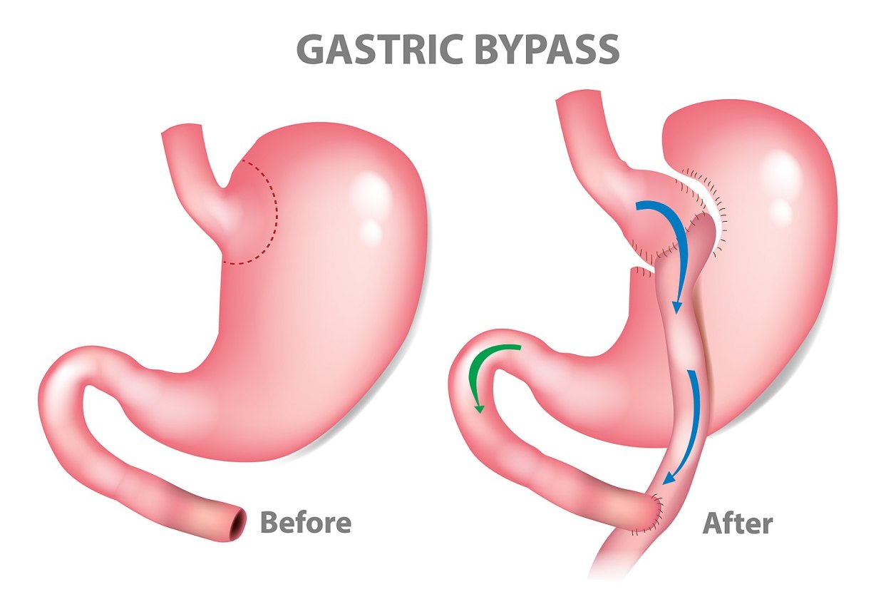
Guest blog: Bariatric Surgery – the safe solution to the metabolic pandemic
A G N Robertson (Twitter: @robertson_a), T Wiggins (@TomWiggins23), F P Robertson, L Huppler (@LucyLucyHuppler), B Doleman, E M Harrison (@ewenharrison), M Hollyman (@misshollyman), R Welbourn
Obesity is the preventable and reversible disease of our lifetime. It is a worldwide health, economic and environmental problem in need of urgent and essential attention, and it has become clear that the world needs more than the traditional recommendations to survive this metabolic pandemic. The traditional advice has been acknowledged for centuries and even more so since the worldwide prevalence of obesity nearly tripled between 1975 and 20161. These lifestyle recommendations include physical exercise, less high calorific food content, balanced meals, optimising portion size, intermittent fasting and so on; we all know them. However, the human race is still falling short of tackling the major public health concern that this disease threatens to be.
Bariatric or weight loss surgery is a surgical sub-speciality which has been evolving since the first procedures of this type in the mid 20th century. Its development has led to the most effective method to achieve long-term weight loss, as well as the additional health benefits weight loss offers as a by-product. However, accessibility to this specialist treatment is limited with only 1% of eligible patients going on to receive bariatric surgery.2Reasons for this limited access are multifactorial, however a considerable factor is thought to be concerns regarding the perceived risks of weight-loss surgery from patients across all populations. We should therefore aim to give our patients the most up to date worldwide risk of mortality of these potentially life-saving procedures.
This month in the BJS, we’ve published the largest meta-analysis asking this question to date – and the findings are pivotal at providing a unanimous international statistic on this discussion. We’ve looked at perioperative mortality rates (inpatient, 30 day and 90 day mortality) of a range of bariatric procedures to include laparoscopic adjustable gastric band (LAGB), sleeve gastrectomy (SG), Laparoscopic Roux-en-Y gastric bypass (LRYGB), one-anastomosis gastric bypass (OAGB), biliopancreatic diversion/duodenal switch (BPD-DS) and other malabsorptive procedures. We’ve included 58 studies in our meta-analysis which has given us information on roughly 3.6 million patients over a 6-year period from worldwide practice. Multiple sources for data were used including administrative datasets, bariatric surgery registries, large scale case series as well as randomised controlled trials (RCTs).
Copied!
Connect

Copyright © 2025 River Valley Technologies Limited. All rights reserved.





.jpg)
