Normal View Dyslexic View
Low presence of intraluminal cancer cells in rectal washout during transanal total mesorectal excision.
21 August 2021
Guest Blog Lower GI
Related articles

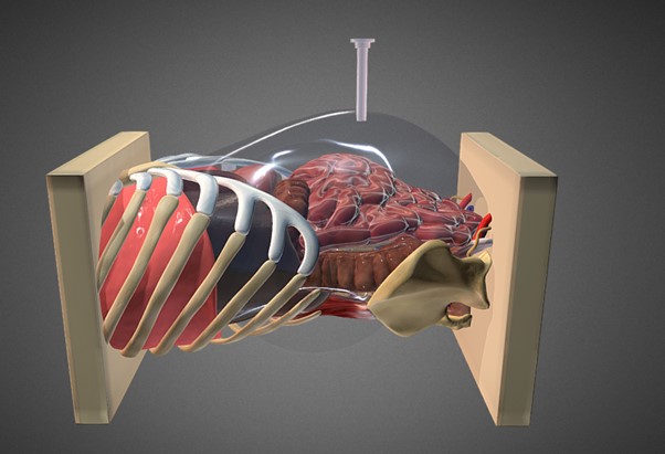
Personalised surgery for the colon cancer patients: new frontiers
B. T. Andersen1,2 A. М. Kazaryan1,2,3,4,5,6 B. V. Stimec7 B. Edwin2,3,8 P. Rancinger1 and D. Ignjatovic2,91 Department of Gastrointestinal Surgery, Østfold Hospital Trust, Grålum, Norway 2 Institute for Clinical Medicine, University of Oslo, Oslo, Norway 3 Interventional Centre, Oslo University Hospital - Rikshospitalet, Oslo, Norway 4 Department of Surgery, Fonna Hospital Trust, Odda, Norway 5 Department of Faculty Surgery, I.M. Sechenov First Moscow State Medical University, Moscow, Russia 6 Department of Surgery N2, Yerevan State Medical University After M.Heratsi, Yerevan, Armenia 7 Anatomy Sector, Teaching Unit, Faculty of Medicine, University of Geneva, Geneva, Switzerland 8 Department of Gastrointestinal and Pediatric Surgery, Oslo University Hospital - Rikshospitalet, Oslo, Norway 9 Department of Digestive Surgery, Akershus University Hospital, Lørenskog, Norway
Introduction
The large bowel is divided into the colon, and the lower part called the rectum and anus. Cancer might develop any place in the large bowel, and usually cancer in the colon or rectum develop from the same type of intestinal cells.
Until the late 1980’s cancer in the rectum typically had a worse prognosis than in the colon. Then, new operative techniques were introduced, and gradually a new understanding of the rectal anatomy developed which led to improvement of the prognosis.
Author’s journey to extraperitoneal left colon surgery.
Tarek S Hany Department of Colorectal Surgery Lancashire Teaching NHS Foundation Trust Preston United Kingdom
From idea to practice.
The transperitoneal (TP) approach to colorectal surgery (both laparoscopic and robotic) has never made sense to me even after many years of performing both techniques. From a colorectal perspective, every important anatomical structure is a retroperitoneal structure. The peritoneum itself is an under-appreciated, under-utilised organ. When trespassing in the peritoneal cavity, the small bowel and omentum present significant unnecessary challenges especially in the obese and those with previous surgery. The whole experience could sometimes feel like trying a knife and a fork for the first time.
Multiport wristed robotic instruments have just made an awkward laparoscopic TP procedure less so. There is a design fault with TP access. There is no doubt, however, that the TP access works in many straightforward operations and this piece is not in any way a call to abandon TP access. However, some 10-25% of operations are responsible for 75-90% of a challenging, long, and painful operation (for patient, surgeon, assistant, and anaesthetist).
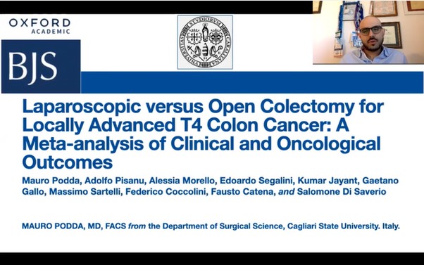
Laparoscopic versus open colectomy for locally advanced T4 colon cancer: a meta-analysis of clinical and oncological outcomes.
Mauro Podda MD FACS, Adolfo Pisanu MD PhD, Alessia Morello MD, Edoardo Segalini MD, Kumar Jayant MD PhD FACS, Gaetano Gallo MD, Massimo Sartelli MD, Federico Coccolini MD, Fausto Catena MD PhD FRCS, and Salomone Di Saverio MD FACS FRCS.
Colon cancer accounts for 9.4% and 10.6% of all tumours in the female and male populations, respectively [Sung H 2020]. Approximately 10-20% of patients diagnosed with colon cancer present with locally advanced tumours penetrating the surface of the peritoneum or directly invading other organs [Brenner H 2014].
This meta-analysis aimed to review the currently available evidence on laparoscopic colon cancer resection, evaluate short postoperative and long-term oncological outcomes after laparoscopic colectomy, and compare the above with conventional open surgery.
We searched medical databases for publications of comparative studies, including randomised controlled trials, prospective cohort studies, and retrospective cohort studies comparing laparoscopic and open surgery as a treatment for T4 locally advanced tumours colon cancers. The searches are up-to-date to April 2021.
Thank you to our strategic partners


.png)


Connect