BJS Academy>Surgical news>A view from the coff...
(1)
A view from the coffee room…use your four basic senses when assessing patients and chronic wounds
Virve Koljonen M.D. PhD.
Department of Plastic surgery
Helsinki University and Helsinki University Hospital
Helsinki, Finland
@plastiikkaope
31 January 2024
Guest Blog General
(1)
Related articles

A view from the coffee room… congress abstracts – good science or bad science?
Virve Koljonen, MD, PhD
Department of plastic surgery
Helsinki University and Helsinki University Hospital
Helsinki, Finland
@plastiikkaope
I vividly remember when attending congresses was in real life only. Today that seems like another reality. I do remember the awkward moments, when I had like one microsecond to decide how to greet my congress acquaintances. Should I greet them with familiarity, or formally, or just casually wave my hand when walking by. As a person coming from a northern country, apart from choosing the right way to greet acquaintances, another custom of the continental Europe is causing me quite a lot of anxiety: air kisses. Which cheek comes first and how many are appropriate, because I don’t want to seem like a stalker, and what if I accidentally belch simultaneously. So many things to consider and be afraid of. Traditionally the success of congresses have been measured by the number of participants and by the number of abstracts 1. This obviously translates to money made by the organizing entity. Before the advent of internet, medical congresses were truly occasions where top scientific innovations were presented, new techniques were introduced, and with a chance to learn from experts 2. Nowadays knowledge is available for anyone at anytime and anywhere. Of course, I will not dismiss the value of face-to-face interactions, that has led to great innovations and is personally meaningful to those participating in such actions. What about the keynote speakers – superstars of their specialty. The more famous the keynote speaker, the more attractive congress is likely to be. In social media, there are events that are called meet-and-greet events. By definition, these events are arranged so that a famous person i.e. influencer can meet and talk to the people. Or is it vice versa actually. Meet-and-greet events are not quite the normal fan event, but almost. Well, to me it sounds a lot like inviting keynote speakers.
A view from the coffee room… Pokémon vs. Predator
Virve Koljonen, MD, PhD
Department of plastic surgery
Helsinki University and Helsinki University Hospital
Helsinki, Finland
@plastiikkaope
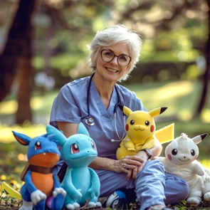
I am a big fan of Pokémon Go. I play it regularly and by that, I mean, every day. The inventiveness of the appearance of Pokémon characters and their witty back stories appeal to me. It is nice to look at a new character and try to find out its real-world counterpart. Further, the structure of the game is to collect as many as possible Pokémon or walking a certain amount of kilometers doing projects within the specified time, perfectly aligning with my competitive personality. Some time ago I was browsing through the medical literature. I am always trying to keep up with new literature, although nowadays it is very difficult. It has been estimated that medical knowledge doubles in just 73 days 1. I do really miss those golden old days when you just did a brisk walk to the library to find what you were looking for. I cannot overestimate my joy when I found out that my favorite leisure time hobby, Pokémon go was employed to expose predator publishers2! Pokémons have helped to reveal that predatory publications have no peer review, nor editing, and what is most choking, not even a reality check2. I am not going the reference these publications, since I feel that the journal gets undeserved glory for including them in the reference list. However, I will walk you through some of these genius publications. For the purposes of this article, I also made AI images in the Pokémon go -style.
A surgical life by Michael G Sarr
I trained at the Johns Hopkins School of Medicine (1972-1976) followed by a surgery residency at the Johns Hopkins Hospital (1976-78 and 1980-1984) including 2 years as a research fellow at the Mayo Clinic (1978-1980) under the direction of Dr Keith Kelly, a truly remarkable surgical scientist. I spent a year as a postdoctoral fellow at Johns Hopkins Hospital (1984-1985) under the tutelage of John Cameron and Gregory Bulkley; both formidable mentors. I was groomed to become the classic academic general surgeon of the times with a primary clinical and research interest in GI surgery and physiology and so I was appointed as an academic surgeon with an NIH-funded laboratory studying GI motility and absorption. My career then spanned the next 37 years at Mayo Clinic with extramural leadership positions in several national and international societies, a 5-year stint as a Director on the American Board of Surgery, Chair of the Department of Veteran’s Affairs Merit Review Committee (1995-1998), and editor of the journal SURGERY for 22 years (2000-2020). If I let my mentors down, it was that I never became Chair of a department of surgery, because I never wanted it. I would never have wanted the administrative responsibilities to interfere with my laboratory pursuits, hands on teaching and clinical interests. All of this is ancient history. What is more important is what follows and hopefully will help young surgeons find their way in our house of surgery. What made you decide to become a surgeon? My dad was a urologist, and as a kid looking at his medical books, I decided I could never be a urologist, but the father of one of my girlfriends was a paediatrician and I really admired him’, so I entered medical school thinking I was going to be a paediatrician ,that is, until I rotated through paediatrics. Two hour rounds 3 times a day and dealing with well-meaning but difficult parents and paediatric nurses who always thought we were barbarians out to hurt the kids (yes, it’s true) was enough to convince me that this was not my calling. I was a very serious student and at Hopkins was being groomed to be an academic physician, so I tried rotations in most specialties. I left the surgery rotation until last, because all I had heard from others was that surgeons were ‘jerks’, were not academic and not scientists. At the end of my third year, I had to do surgery and after 2 weeks, loved it. I enjoyed the work, the challenges, and the surgical personalities, but how could I be an academic as a surgeon? Then I met a several surgeon scientists (there were quite a few at Hopkins) who ran laboratories, were great teachers, and were exciting. From then on, I knew that surgery was my calling.
Copied!
Connect

Copyright © 2026 River Valley Technologies Limited. All rights reserved.




.jpg)
